Meaning of Histology
Histology is the study of the microscopic structure of tissues. It helps us understand how the body is built at the smallest level — what our cells and tissues look like, how they work, and how diseases affect them.
In simple terms, histology is the science of “seeing what the naked eye cannot see.”
For students of Complementary and Alternative Medicine (CAM), histology is important because it provides a biological foundation for understanding how herbs, nutrition, and therapies act on body tissues
2. The Origin and Evolution of Histology
Histology has grown through many centuries of discovery. It started with early scientists trying to understand the human body through dissection, and it advanced when microscopes were invented.
Here’s a brief timeline of how histology evolved:
Ancient Period (Before Microscopes)
Thinkers like Hippocrates and Aristotle described body structures but could only see what was visible to the naked eye.
Anatomy was mainly studied through dissection, without knowledge of cells or tissues.
Renaissance Era (1500s)
Andreas Vesalius (1514–1564) improved human anatomy studies by dissecting human cadavers and correcting earlier errors.
However, tissues were still studied without magnification.
17th Century – The Birth of Microscopy and Histology
Around 1590, Zacharias Janssen and his father built the first compound microscope, allowing magnified viewing of small objects.
Robert Hooke (1665) used this microscope to observe thin slices of cork. He saw tiny box-like structures which he called “cells” — the beginning of cell biology.
Antonie van Leeuwenhoek (1674) made his own simple microscope with powerful lenses. He was the first to see living cells, including blood cells, sperm cells, and bacteria.
18th and 19th Centuries – Foundation of Modern Histology
Scientists began to prepare thin slices of tissues and use dyes (stains) to make the structures more visible.
Marcello Malpighi (1628–1694) is known as the “Father of Histology.” He studied the lungs, liver, and kidneys using an early microscope.
Theodor Schwann and Matthias Schleiden (1838–1839) proposed the Cell Theory, which states that:
1. All living things are made up of cells.
2. Cells are the basic units of life.
3. New cells come from pre-existing cells.
This theory changed biology forever and made histology a key part of medicine.
20th Century – The Rise of Advanced Microscopy
Electron microscopes were developed in the 1930s, allowing scientists to see cell structures at very high magnification — even smaller than what light microscopes could show.
Histology became essential in medicine, pathology, and research.
3. The Relationship Between Histology, Anatomy, and Pathology
– Anatomy looks at organs and structures visible to the naked eye.
– Histology focuses on tissues and cells that make up those organs.
– Pathology studies how diseases cause changes in these tissues.
So, histology is the bridge between anatomy and pathology — it helps explain how normal structures become abnormal in disease.
4. Introduction to Microscopy
The microscope is the most important tool in histology. It allows scientists and students to observe the fine details of tissues and cells.
A. Types of Microscopes
1. Light Microscope (Optical Microscope)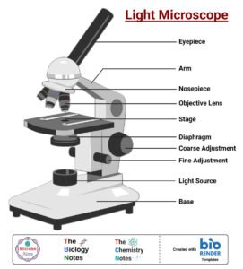
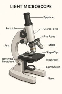
Uses visible light to illuminate the specimen.
Can magnify up to about 1000 times.
Used for viewing stained tissue sections in most histology labs.
Example: observing epithelial cells, muscle fibers, or blood cells.
2. Electron Microscope
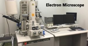
![]()
Uses a beam of electrons instead of light.
Can magnify up to a million times — showing very fine details of cell structures.
Two main types:
Transmission Electron Microscope (TEM): shows internal structures of cells (organelles, membranes).
Scanning Electron Microscope (SEM): shows surface details of tissues in 3D.
3. Fluorescence Microscope (optional advanced type)
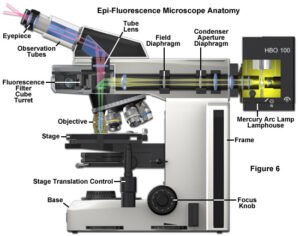
Uses special dyes that glow under ultraviolet (UV) light.
Useful in detecting specific molecules or diseases in tissues.
5. Parts and Functions of a Light Microscope (Simplified)
Eyepiece (ocular lens): where you look through.
Objective lenses: provide magnification (usually 4x, 10x, 40x, 100x).
Stage: platform that holds the glass slide.
Condenser: focuses light on the specimen.
Mirror or light source: provides illumination.
Coarse and fine focus knobs: help bring the image into clear view.
6. Care and Handling of the Microscope
Carry the microscope with both hands (one under the base, one on the arm).
Start with low power before switching to high power.
Always clean lenses with lens paper, never with tissue or cloth.
Do not use direct sunlight as a light source.
Keep the microscope covered when not in use to prevent dust.
7. Importance of Microscopy in Complementary and Alternative Medicine (CAM)
Helps in understanding how natural therapies work on tissues.
Enables observation of tissue damage or healing processes.
Supports herbal research — e.g., studying the effects of plant extracts on liver or skin cells.
Enhances diagnostic ability in natural health practice
8. Summary
Histology = study of microscopic tissues.
Originated after the invention of the microscope in the 1600s.
Robert Hooke discovered cells; Leeuwenhoek observed living ones.
Malpighi is the Father of Histology.
Light microscopes are used for general tissue observation.
Electron microscopes reveal detailed cell structures.
Histology connects structure (anatomy) with disease (pathology) — essential for understanding health and healing.
The video for this module is credited to DaVinci Academy. It is embedded directly from their official YouTube channel for educational purposes only.