Specialized Connective Tissues II – Cartilage
1. Introduction
Cartilage is a tough but flexible type of specialized connective tissue that provides support, shape, and cushioning to various parts of the body. It serves as a smooth surface for movement at joints and forms the framework for some structures such as the ear, nose, and respiratory passages. Unlike bone, cartilage does not contain blood vessels or nerves, which makes it slower to heal when damaged.
2. Characteristics of Cartilage
Cartilage has a firm, rubbery matrix that gives it strength and flexibility.
It contains specialized cells called chondrocytes that lie in small spaces called lacunae.
It is covered by a connective tissue membrane known as the perichondrium, except at joint surfaces.
It is avascular (lacks blood supply) and receives nutrients through diffusion from surrounding tissues.
3. Functions of Cartilage
Provides smooth surfaces for joint movement.
Supports soft tissues and maintains the shape of structures such as the ear and nose.
Serves as a model for bone formation during development (endochondral ossification).
Acts as a cushion and shock absorber in joints.
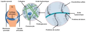
4. Composition of Cartilage
Cartilage is made up of three main components:
Cells: Chondroblasts (young cells) and chondrocytes (mature cells).
Fibers: Collagen and elastic fibers for strength and flexibility.
Matrix: A firm, gel-like substance that contains water, proteins, and sugars.
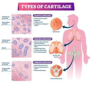
5. Types of Cartilage
A. Hyaline Cartilage
Most common type of cartilage.
Has a smooth, glassy appearance with fine collagen fibers.
Function: Provides support with some flexibility; reduces friction between bones.
Location: Ends of long bones (articular cartilage), trachea, nose, and larynx.
B. Elastic Cartilage
Contains many elastic fibers that make it more flexible than hyaline cartilage.
Function: Maintains shape while allowing flexibility.
Location: External ear (pinna), epiglottis, and auditory tube.
C. Fibrocartilage
Contains thick bundles of collagen fibers, making it the strongest type.
Function: Provides toughness and resists compression.
Location: Intervertebral discs, pubic symphysis, and knee joints (menisci).
6. Cells of Cartilage
Chondroblasts: Actively produce cartilage matrix; found near the surface under the perichondrium.
Chondrocytes: Mature cartilage cells located within lacunae; maintain the matrix.
7. Growth of Cartilage
Cartilage grows in two main ways:
Appositional growth: New layers are added to the surface by chondroblasts in the perichondrium.
Interstitial growth: Chondrocytes divide and secrete new matrix within the cartilage, increasing its size from inside.
8. Perichondrium
The perichondrium is a dense layer of connective tissue surrounding most cartilage.
Functions:
Supplies nutrients to the cartilage through diffusion.
Contains cells that help in cartilage growth and repair.
9. Clinical Relevance
Cartilage injuries heal slowly because of lack of blood supply.
Degeneration of articular cartilage can cause arthritis.
In Complementary and Alternative Medicine, supporting cartilage health involves nutrition rich in collagen, vitamin C, and natural anti-inflammatory herbs to protect joints and enhance flexibility.
10. Summary
Cartilage is a specialized connective tissue made up of chondrocytes in a firm, flexible matrix.
There are three main types: hyaline, elastic, and fibrocartilage.
It provides support, flexibility, and smooth surfaces for movement.
Because it lacks blood supply, cartilage heals slowly.
Maintaining healthy cartilage is important for joint function and mobility, and CAM therapies emphasize diet and natural remedies that support joint and cartilage regeneration.
Video Credits
The educational videos in this course are credited to the following creators:
All videos are embedded directly from YouTube for educational purposes and remain the property of their respective creators.